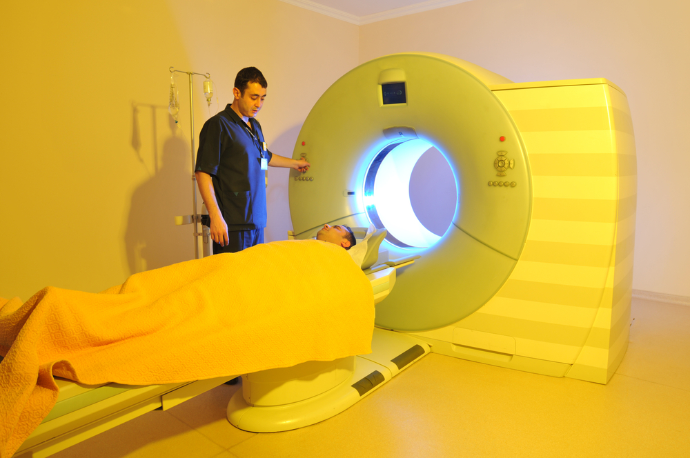
 A recent study led by Johns Hopkins’ researchers suggests it is possible to distinguish cancerous from noncancerous cells using an MRI based on sugar molecules, avoiding invasive biopsies. This MRI technique was tested both in mice and test tube-grown cells, and the results were published in Nature Communications.
Imaging tests like CT scans or mammograms can detect the presence of tumors but cannot fully render a cancer diagnosis, as a biopsy is usually required to confirm imaging results. This study revealed that non-invasive MRIs can make biopsies more effective or even replace them through detection of telltale sugar molecules shed by the outer membranes of cancerous cells.
“We think this is the first time scientists have found a use in imaging cellular slime. As cells become cancerous, some proteins on their outer membranes shed sugar molecules and become less slimy, perhaps because they’re crowded closer together. If we tune the MRI to detect sugars attached to a particular protein, we can see the difference between normal and cancerous cells,” said study author Jeff Bulte, a professor at the Institute for Cell Engineering at the Johns Hopkins University School of Medicine.
[adrotate group=”1″]
Bulte’s research was based on recent findings that indicated glucose could be detected through a fine-tuned MRI technique. Bulte’s research team managed to compare MRI readings from proteins known as mucins (a family of proteins known for its high molecular weight) with and without sugars attached, assessing how the signal was modified. This signal was investigated in 4 types of lab-grown cancer cells, with results showing lower levels of mucin-attached sugars in cancer cells, when compared to normal cells.
Xiaolei Song, lead author, explained: “The advantage of detecting a molecule already inside the body is that we can potentially image the entire tumor.”
More testing is required to prove this technique could be valuable for human cancer diagnosis and the research team will continue its efforts to understand what types of tumors this method can be used on. However, the prospect of being able to accurately diagnose cancer in its earlier stages, with the prospect of eventually doing so without the use of biopsies, could lead to significantly improved cancer patient outcomes as well as reduced healthcare costs.


