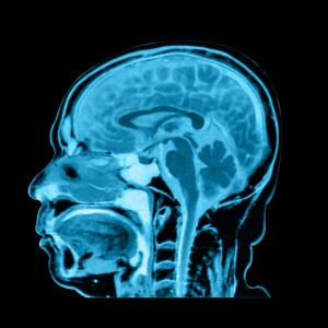
 A recent study published in the ACS Applied Materials & Interfaces journal found that green tea can improve the quality of MRI images.
In recent years, green tea has been found to be a beneficial health supplement, thanks to polyphenols like flavonoids and catechins that function as antioxidants. Green tea is also used in some foods and even as a skin moisturizer. According to researchers, green tea can also improve the image quality of MRIs, as laboratory studies have successfully used it to image cancer in mice models of tumors.
The research team led by Dr. Sanjay Mathur noted the potential utility of nanoparticles and iron oxide to improve bio-medical imaging. However, nanoparticles have associated disadvantages: they tend to cluster together quite easily and need assistance to get to their destinations in the body.
In order to address these issues, scientists have tried to attach natural nutrients to the particles to help guide them. What Dr. Mathur’s team evaluated was if green tea compounds could actually play this role, since previous research suggested that green tea has both anti-cancer and anti-inflammatory properties.
[adrotate group=”1″]
Through a simple and single-step process, scientists coated iron-oxide nanoparticles with catechins, green-tea compounds, and administered them to mice suffering with cancer. The resulting MRIs showed that the new imaging agents could bind to tumor cells, resulting in a stronger contrast from non-tumor cells surrounding that area. Researchers concluded that the catechin-coated nanoparticles might actually be promising candidates to improve image in MRIs and could be used for other related applications.
Read More News About MRI
A recent study entitled “Training and Standards for Performance, Interpretation, and Structured Reporting for Supplemental Breast Cancer Screening“ led by Wendie A. Berg and published in the American Journal of Roentgenology (AJR) found that ultrasound lags behind MRI when it comes to screening for supplemental breast cancer. (read more)
A recent large-scale study has reported that targeted biopsy with new fusion technology, which joins magnetic resonance imaging (MRI) and ultrasound, was found to be more efficient in identifying high-risk prostate cancer when compared to standard biopsy. The study, entitled “Comparison of MR/Ultrasound Fusion-Guided Biopsy With Ultrasound-Guided Biopsy for the Diagnosis of Prostate Cancer,” published in the renowned journal JAMA and conducted at the National Institutes of Health (NIH), from 2007 to 2014, enrolling a total of 1,003 individuals. (read more)


