
 A research team from the University of California, San Diego School of Medicine has published their latest research in the Prostate Cancer and Prostatic Disease journal, whereby they describe a new imaging technique that can significantly improve prostate cancer imaging, resulting in improved treatment outcomes.
“This new approach is a more reliable imaging technique for localizing tumors. It provides a better target for biopsies, especially for smaller tumors,” study author Rebecca Rakow-Penner, MD, PhD, a research resident in the Department of Radiology, said in a news release.
This novel technique can also aid in treatment planning and image presentation, explained study author David S. Karow, MD, PhD, assistant professor of radiology at UC San Diego. “Doctors at UC San Diego and UCLA now have a non-invasive imaging method to more accurately assess the local extent of the tumor and possibly predict the grade of the tumor, which can help them more precisely and effectively determine appropriate treatment”, he added.
Usually, for prostate cancer detection, specialists use contrast enhanced magnetic resonance imaging (MRI), which consists of an intravenously injected die that upon higher blood flow (a common characteristic of tumors) allows the distinction between normal tissue and cancerous one.
However, many times this technique is not sensitive enough to correctly detect tumors, leading experts to use diffusion MRI measures, which can detect different tissue’s density (cancer tissues are denser than healthy ones). Once more, this technique has its flaws and can suffer from magnetic field artifacts that lead to erroneous tumor localization, which is of particular relevance when surgeons need to excise a tumor and understand potential spread, or upon radiation treatment planning, which requires a precise knowledge of tumor localization.
[adrotate group=”1″]
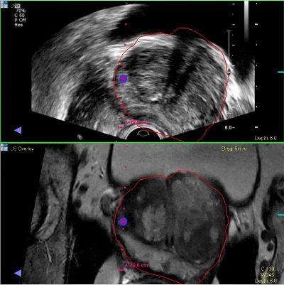
The new technique developed in this study is called restriction spectrum imaging-MRI (RSI-MRI), and can adjust for magnetic field distortions and focus upon water diffusion within cancer cells. This allows an accurate image to better localize the tumor.
“Prostate cancer can often be an indolent disease, where a patient may only require surveillance rather than aggressive surgery,” study author Christopher J. Kane, MD, professor of urology at UC San Diego, explained.
“If by imaging we could predict the tumor grade,” added Robert Reiter, MD, professor of urology at UCLA, “we may be able to spare some patients from prostate resection and monitor their cancer with imaging.”
