
 In a study entitled “Inter-observer variability of clinical target volume delineation in radiotherapy treatment of pancreatic cancer: a multi-institutional contouring experience” published in the Radiation Oncology journal, a team of researchers designed an observational multi-institutional study to evaluate the inter-observer variability in clinical target volume (CTV) delineation among different radiation oncologists in radiotherapy treatment of pancreatic cancer (PC).
Patients with pancreatic adenocarcinoma usually have a poor prognosis, and both chemotherapy and radiotherapy (RT) have been used for unresectable disease as well as in the adjuvant setting to help improve survival.
A large percentage of PC patients die from local uncontrolled disease, and lymph node metastases have proven to be an important prognostic factor associated with a significantly higher rate of both local and distant recurrences.
During radiotherapy planning, local control is an important treatment end-point with different volumes needing to be considered.
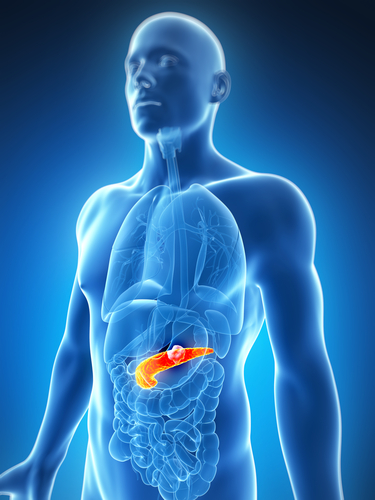
CTV contains the gross tumor volume, or what can be seen, palpated, or imaged from the tumor, plus a margin for sub-clinical disease spread, which cannot be fully imaged. CTV measurement is important because this volume must be adequately treated to achieve a cure, however, it can be quite challenging to measure because it cannot be accurately defined for an individual patient.
It is still not established whether elective nodal irradiation (ENI) should be performed to treat regional nodal metastases in patients with PC.
In this study, researchers analyzed two different cases of pancreatic cancer treated by postoperative and preoperative RT, including parameters such as clinical history, diagnostics, and planning CT imaging.
Participants were asked to delineate CTVs according to their skills and knowledge, with the total volume, craniocaudal, laterolateral, and anteroposterior diameters calculated to quantify interobserver variability of CTVs delineations.
Moreover, the Dice Similarity Index (DSI) was used to evaluate the spatial overlap accuracy of the different CTVs and compare it with the CTVs of a national reference Centre.
[adrotate group=”1″]
Researchers observed that among the 18 radiation oncologists from different Institutes that participated, less variability was observed for elective CTV rather than boost CTV, two criteria used in CTV definition and delineation.
The estimated clinical volumes (CV) were 28.8% and 20.0% for the elective CTV, in adjuvant and neoadjuvant cases, respectively, with a mDSI value of 0.68 for elective CTV in both cases. Additionally, a CV of 42.4% and 63.8% with a mDSI value of 0.44 and 0.52, were calculated for boost CTV in both cases, respectively.
These results show an agreement in both post-operative and pre-operative scenarios, demonstrating that the availability of national reference criteria has produced reliable results.
In the future, the authors propose that additional strategies to increase the agreement in elective CTV and reduce variability in boost CTV delineation could be obtained by implementing the routinely use of intravenous contrast-enhanced planning CT scan and through promotion of a collaborative effort between multidisciplinary programs involving radiologists and nuclear physicians.
