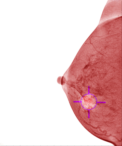
 The University of California, Irvine, has established a partnership with Royal Philips in order to study spectral breast imaging and its effects in the improvement of measurement of breast density. The investigators expect to provide physicians with more accuracy in the method to determine the risk of developing breast cancer, as well as effectively monitoring the disease.
The study that is going to be initiated from the collaboration is focused on MicroDose SI mammography spectral imaging technology from Royal Philips, which was created to fulfill the medical need for more accurate methods of breast cancer diagnosis. There is great concern regarding regarding the low accuracy of mammography for high density breasts in diagnosing the disease, and MicroDose SI is expected to improve the quality of imaging and consequently the accuracy of the results.
[adrotate group=”1″]
“Through this study, UC Irvine and Philips are looking to set an industry standard for objectively measuring breast density,” explained the CEO Imaging at Philips, Gene Saragnese, in a press release. “While this doesn’t exist today, it will be increasingly critical as we move toward further personalizing breast cancer screening, and enabling patients to become more engaged in their own care.”
“With the combination of our MicroDose SI technology and leading imaging experts at UC Irvine, we can determine if spectral breast imaging can help provide more definitive diagnoses, at low radiation dose, to better help patients and clinicians in the fight against breast cancer,” added Saragnese.
MicroDose SI is the first spectral imaging application from Philips and includes a feature of Spectral Breast Density Measurement, which uses photon counting technology to register spectral data of the adipose and fibroglandular tissue at the same time exposing the patient to only one low dose mammogram. With this information about volumetric breast density, the device may redefine risk evaluation and personalized care.
Higher breast density increases the risk of suffering from breast cancer, as women who have 75% or more dense tissue are four to six times more likely to develop the disease than women with little or no dense tissue. On the other hand, breast density also makes it harder for breast cancer to be detected during a mammography. Given that, more accurate evaluations may help radiologists to provide personalized breast cancer screenings and therapy monitoring if necessary.
“One of the biggest challenges for us has been the lack of quantitative standards, which makes it very difficult to prove the accuracy of breast density measurements,” added the lead researcher in the study, Sabee Molloi, PhD, who is a professor of radiological sciences at the University of California, Irvine School of Medicine. “Leveraging Philips’ spectral imaging technology, this study will help us validate breast density measurements and set industry standards, which can help enhance the quality of diagnoses and treatment.”
The study will be initiated with the evaluation of the current application of spectral breast density, through the examination of 40 post-mortem breasts and a comparison with the outcomes from previous chemical analysis. The university and the company expect the study to be completed within a year or two.


