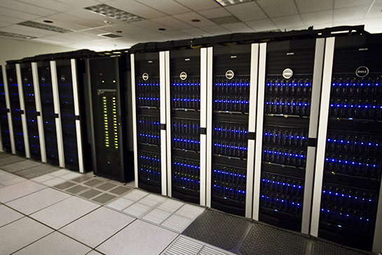
A research project at the MD Anderson Cancer Center in Houston focused on tracking how much radiation is being delivered through the MRI-linac is using simulations on the Texas Advanced Computing Center (TACC)’s Lonestar supercomputer to model radiation in a magnetic field, developing a technique that will facilitate the safe use of the MRI-linac and enable more effective cancer treatment. The MRI-linac offers a potentially significant improvement over current cancer treatment technology by uniting radiation therapy with magnetic resonance imaging (MRI).
Medical researchers and physicians are obliged to walk a delicate line, delivering just enough radiation to kill tumors while sparing surrounding healthy tissue. “By running the supercomputer simulations, our goal is to  understand how to properly use ionization chambers in the presence of a magnetic field. The results of our research will facilitate safe use of the MRI-linac systems, which in turn will be used to treat cancer patients more effectively,” says Michelle Mathis, a Postdoctoral Fellow at MD Anderson Cancer Center and medical physics researcher for the MD Anderson dosimetry team in a TAcc release. “Precise knowledge of dose is critical to effective radiation treatment.”
“Different tumors need different doses to be killed,” Dr. Mathis observes. “Our work focuses on gaining a better understanding of how to precisely calibrate the new MRI-linac system so that the appropriate amount of radiation is delivered to the cancer tumor while healthy tissue is spared.”
Delivering the Right Dose Using Ionization Chambers
Radiation therapy is typically administered through a linear accelerator, or linac. Inside the linac, electrons are accelerated through an electric field, until they’re traveling at nearly the speed of light. Once the electrons reach this speed they hit a target, creating radiation. The radiation is then shaped in the linac, and sprayed down to target the tumor in a patient’s body.
“Cancer is a global issue, so it’s nice to take a holistic approach in improving technology to treat it — ultimately saving more lives and ensuring a better overall quality of life for those affected by cancer.” Dr. Mathis notes.
Medical physicists use a type of radiation detector called an ionization chamber to ensure that the radiation dose delivered is accurate. Ionization chambers are a necessary part of dose measurement, allowing medical physicists to calibrate the linac. However, integrating radiation therapy and MRI into a single system adds a magnetic field, which adds a new layer of complication to the performance of ionization chambers.
“In order to adjust for the presence of a magnetic field, we are developing correction factors so that ionization chambers can be used to calibrate the amount of radiation delivered by the new MRI-linac system,” Dr. Mathis explains
Since only one prototype of the MRI-linac system is available, Dr. Mathis performs supercomputer simulations to model radiation in a magnetic field. The MD Anderson dosimetry team is using a total of 250,000 hours on Lonestar and will continue to run simulations until they can develop correction factors for 16 different ionization chambers.
The TACC notes that current radiation treatment relies on imaging from computed tomography (CT) scans, which are taken prior to treatment to determine the tumor’s location. This method works fairly well if the tumor lies in an easily detectable and immobile location.
[adrotate group=”1″]
However, if a patient has lung cancer and will soon undergo radiation treatment. CT scans don’t provide real-time imaging of the tumor and with the tumor constantly moving due to the patient’s breathing. The doctor might end up irradiating a large portion of the lung unnecessarily, which could destroy the tumor, but could also lead to a lower quality of life due to effects like difficulty breathing and reduced mobility.
As part of a checks and balances system, one research project at the MD Anderson Cancer Center in Houston focuses on dosimetry — tracking how much radiation is being delivered through the new radiotherapy device.
The Texas Advanced Computing Center (TACC) at The University of Texas at Austin provides the necessary support to Mathis’ research to run these simulations. Through the UT System Research Cyberinfrastructure (UTRC), an initiative that allows any of the 15 University of Texas System institutions access to TACC’s supercomputing resources, the team runs simulations on Lonestar, a powerful, high performance computing and remote visualization resource.
“Using Lonestar, we are able to simulate the effect of many variables on the ionization chamber readings, which will allow us to precisely calculate radiation dose,” Dr. Mathis explains.
 “Many institutions have their own clusters, however, their machines aren’t as powerful or well-maintained as TACC’s,” says John Fonner, a research associate in TACC’s Life Sciences Computing Group. “And because TACC staff are professionals in the advanced computing world, they can provide the technical expertise that other institutions can’t, allowing researchers to take their projects to the next level.”
The TACC notes in a release that the UTRC Project provides a strong foundation for advances in current and future research efforts across UT System. The combination of high bandwidth access, persistent data storage, computational capabilities, and the expertise of UT System institution researchers have incredible potential and keep UT System at the forefront of science and discovery.
The collaboration between UT System and The Texas Advanced Computing Center (TACC) leverages advanced computing and visualization resources via the Lonestar system, data storage and collections resources via UT Data Repository (UTDR), as well as, TACC support services to enable researchers to learn to use UTRC resources effectively. TACC staff members assist research teams in properly employing the systems, software, and expertise necessary to foster scientific innovation and are dedicated to providing the research & development, consulting, and training required so users can take full advantage of TACC resources, services, and training programs that are contributing to the development of the next generation of researchers. TACC’s talented staff members strive to provide the technology, computing, and visualization assistance that empower UT System’s computational research community to impact science and change the world.

The MD Anderson dosimetry team is using a total of 250,000 hours on Lonestar and will continue to run simulations until they can develop correction factors for 16 different ionization chambers. “By running the supercomputer simulations, our goal is to understand how to properly use ionization chambers in the presence of a magnetic field,” Dr. Mathis says. “The results of our research will facilitate safe use of the MRI-linac systems, which in turn will be used to treat cancer patients more effectively.”
The new MRI-linac has not been released, but the pieces are starting to come together. Several institutions in the consortium have recently been installing initial components (more on this below). Dr. Mathis’ dosimetry team will continue working to better understand the system and ensure it functions as intended.
Cancer is a global issue, so it’s nice to take a holistic approach in improving Dr. Mathis observes, “Technology to treat it — ultimately saving more lives and ensuring a better overall quality of life for those affected by cancer.”
Another research group, a joint effort of
Elekta — a Swedish company that provides radiation therapy, radiosurgery, related equipment and clinical management for the treatment of cancer and brain disorders — and Philips Research Consortium on MRI-Guided Radiation Therapy, are tackling this issue by developing a potentially significant improvement over current technology — the MRI-linac.
Elekta and Royal Philips announced recently that The Institute of Cancer Research, London, a world-leading cancer research institution, working with its clinical partner The Royal Marsden NHS Foundation Trust, will join the Elekta MR Linac Research Consortium, a group with a mission to develop an integrated magnetic resonance imaging (MRI) guided radiation therapy system. Uniting an advanced digital linac with an MRI system would greatly enhance real-time visualization of cancer targets during the delivery of therapeutic radiation.

In collaboration with clinical partners, Philips and Elekta will seek to merge advanced Magnetic Resonance Imaging ( MRI, picture on the left) with precision radiation therapy (picture on the right) in a single MRI-guided radiation therapy system. This will enable doctors to achieve exceptional soft tissue imaging during radiation therapy and to adapt treatment delivery in real-time for extremely precise cancer treatments
 “The addition of The Institute of Cancer Research (ICR) to the consortium further strengthens this growing partnership of clinical centers,” said Niklas Savander, Elekta President and CEO. “The ICR is a prominent cancer research institute and has contributed significantly in this arena for over a century. Its unique partnership with The Royal Marsden and ‘bench-to-bedside’ approach allows the ICR to deliver exceptional results. All of these factors make the ICR and The Royal Marsden a perfect fit as we advance MRI-guided radiotherapy.”
 “The development of MRI-guided radiation therapy establishes a new vision for clinical radiation oncology for the treatment of cancer,” observes Prof. Uwe Oelfke, MCCPM, FInstP, and Head of the Joint Department of Physics at The Institute of Cancer Research, London and The Royal Marsden NHS Foundation Trust, in a release. “The translation of this novel technical approach into clinical practice faces a number of challenges ideally addressed by a group of world leading radiotherapy centers, each contributing with its specific expertise. Being a member of the consortium is a prerequisite to shape this exciting future of radiation therapy.”
“Our current Cancer Research UK-funded research program on adaptive and image guided therapy,” says Dr. Oelfke, “together with the activities on MRI within our Cancer Research UK imaging center, are an excellent research environment to make leading contributions to the joint efforts of the consortium. The ICR’s initial research focus for MRI-guided radiation therapy will be on pelvic malignancies, where the excellent soft tissue contrast of MR images directly before or during treatment will substantially enhance treatment options and quality.”
Last year, Elekta and Royal Philips Electronics announced that The Netherlands Cancer Institute-Antoni van Leeuwenhoek Hospital (NKI-AVL, Amsterdam, the Netherlands) had signed an agreement to join a research group to advance development of ground-breaking image-guided treatment technology for cancer care. The technology merges radiation therapy and magnetic resonance imaging (MRI) technology in a single system. NKI-AVL was the third member of the now six-member research consortium, pooling efforts of leading radiation oncology centers and clinicians researching state-of-the-art radiation therapy and MRI in a single system. The consortium currently includes the University Medical Center Utrecht (Utrecht, the Netherlands), The University of Texas MD Anderson Cancer Center (Houston, Texas), The Netherlands Cancer Institute-Antoni van Leeuwenhoek Hospital (Amsterdam, the Netherlands), Sunnybrook Health Sciences Centre (Toronto, Ontario) and The Froedtert & Medical College of Wisconsin Cancer Center (Milwaukee, Wisconsin).
 “MRI-guided radiation therapy has the promise to become a highly targeted treatment for cancer,” says Gene Saragnese, CEO Imaging Systems at Philips Healthcare.. “The realization of this promise requires the collaboration between leaders in interventional imaging and radiation therapy delivery, as well as clinical innovators in oncology. We are convinced that the deep clinical knowledge and expertise of the Institute of Cancer Research will be a strong asset to the growing research consortium.”
Uniting state-of-the-art MRI with a cutting edge radiation therapy system — thus creating an MRI-guided radiation therapy system — will provide physicians with exceptional images of a patient’s soft tissues and tumor during radiation therapy. This breakthrough innovation also aims to permit clinicians to adapt treatment delivery in real time for the most precise cancer treatments possible.
“MRI has steadily revolutionized healthcare since its introduction nearly three decades ago, giving clinicians unparalleled views of soft tissues and pathology. Merging this diagnostic capability with the capacity to also treat disease in the same frame of reference could dramatically improve cancer management,” said Tomas Puusepp, who was Elekta President and CEO, at the time. “The other consortium members at Elekta, Philips, University Medical Center Utrecht and MD Anderson are delighted that NKI-AVL – an internationally renowned medical center – has joined us in this important effort.”
 Prof. Marcel Verheij, Head of the Radiotherapy Division at NKI-AVL, commented that: “MRI-guided radiotherapy allows optimal imaging and will therefore improve the accuracy of our treatment delivery. Building on our experience with Cone Beam CT-guidance we are highly motivated to collaborate within the research consortium and contribute to the implementation of MRI-guided adaptive radiotherapy… This research exemplifies the essential role that imaging plays in the development of more targeted treatments for cancer.”
Philips Healthcare’s Gene Saragnese, noted that “The Netherlands Cancer Institute-Antoni van Leeuwenhoek Hospital has played a crucial role in the software development for CT-guided radiation therapy a decade ago, and this expertise complements the skills that we already possess in this research consortium.”
Prior to establishment of the research consortium, Elekta, Philips and the University Medical Center Utrecht built and tested a prototype system that integrates a linear accelerator and a 1.5 Tesla MRI system. The success of these efforts enabled the project to move to the next phase of development and testing by the growing select group of consortium partners.
The new systems uniting radiation therapy and magnetic resonance imaging (MRI) will allow physicians to view the tumor in real-time and in high detail during treatment. It also permits physicians to adapt the radiation treatment during the procedure, sparing healthy tissue and reducing side effects.
The research consortium comprises corporations, hospitals, universities, and research institutions worldwide, all of which contribute the distinct research components necessary to develop this technology.
Sources:
Texas Advanced Computing Center
MD Anderson Cancer Center
Elekta
Royal Philips Electronics
Elekta MR Linac Research Consortium
The Institute of Cancer Research, London
Image Credits:
MD Anderson Cancer Center
Texas Advanced Computing Center
Imperial College, London


