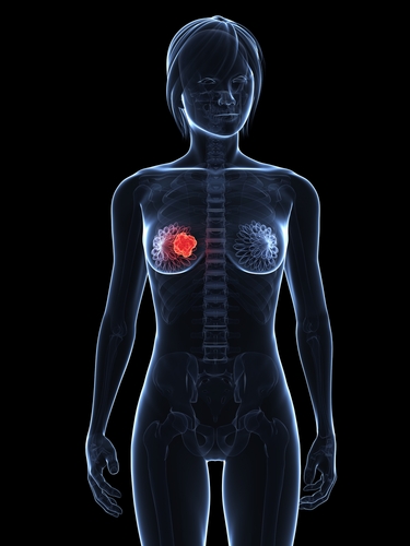
 Results from the study entitled “Molecular Breast Imaging at Reduced Radiation Dose for Supplemental Screening in Mammographically Dense Breasts”, were recently published in the American Journal of Roentgenology by Mayo Clinic researchers.
The performance of digital screening mammography is reduced in women with mammographically dense breast tissue, who constitute approximately 50% of the screening-aged population. According to breast density inform legislation, women with dense breasts undergoing mammography may benefit from supplemental screening, however at the moment, there is a lack of consensus about the best supplemental imaging technique and the balance of its benefits and harms.
Deborah Rhodes and colleagues at the Mayo Clinic in Rochester, examined the diagnostic performance of supplemental screening molecular breast imaging (MBI) in 1,651 women with mammographically dense breasts after system modifications to permit radiation dose reduction.
A total of 1,585 patients completed the reference standard from May 2009 to March 2012. Of these, 21 were diagnosed with cancer using either pathology findings (2.5%), negative imaging findings (96.1%), clinical examination (0.1%), and patient interview (1.3%).
Based on their study results, researchers determined that upon using supplemental MBI with mammography cancer detection rates increased from 3.2 to 12.0. Furthermore, supplemental MBI was found to increase invasive breast cancer detection rate from 1.9 to 8.8. “Approximately 80% of cancers seen only on MBI were invasive, suggesting that MBI is not selectively detecting clinically unimportant cancers. Eighty-two percent of the invasive cancers seen only on MBI were node negative, suggesting that MBI contributes to early detection of clinically important cancers”, the authors write in their study. “MBI also detected larger and node-positive invasive cancers that were mammographically occult, suggesting a role for MBI in identifying cancers masked on repeated mammographic screenings by dense breast parenchyma”, they add.
[adrotate group=”1″]
When assessing detection sensitivity, specificity and positive predictive value of the biopsies performed (PPV3), researchers found that for mammography alone, sensitivity was of 24%; specificity of 89%; and PPV3 25%. Combining MBI with mammography revealed a detection sensitivity of 91%, specificity of 83%; and PPV3 of 28%. In terms of recall rate, researchers found mammography alone had a rate of 11.0%, which increased upon MBI addition (17.6%).
According to the authors, results from this study strongly indicate that when added to screening mammography, molecular breast imaging with a reduced radiation dose (effective dose 2.4 mSv), can add an incremental cancer detection rate of 8.8 per 1000 women with mammographically dense breasts.


